


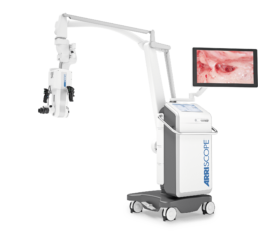
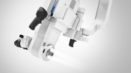
Native Digital 3DSurgical Microscopy
The ARRISCOPE is the world’s first high-definition, fully digital surgical microscope for stereoscopic viewing. It allows for live image augmentation and bringing together all relevant information directly into the surgeon’s field of view. This transforms the ARRISCOPE into a pivotal visualization system in the OR.
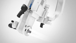
Native Digital 3D Imaging
The ARRISCOPE takes microsurgical imaging to the next technological level. Its all-digital 3D imaging chain provides crystal-clear high-resolution images and videos. Both, LED illumination and the entire imaging chain, are manufactured by ARRI and are therefore optimally coordinated.
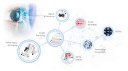
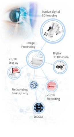
The ARRISCOPE takes microsurgical imaging to the next technological level. Its all-digital 3D imaging chain provides crystal-clear high-resolution images and videos. Both, LED illumination and the entire imaging chain, are manufactured by ARRI and are therefore optimally coordinated.


See whatreally matters
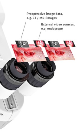
The digital 3D binoculars are the core element for visualizing key information directly in the surgeon‘s field of view. The live image, as well as preoperative image data and any other video signals, can be selected for viewing individually or simultaneously. The surgeon can remain immersed completely in the procedure and call up the precise data he needs at any time.
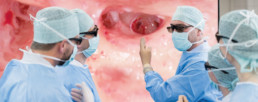
A New Benchmark in Clinical EducationAssist Mode
A New Benchmark in Clinical EducationAssist Mode
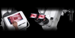
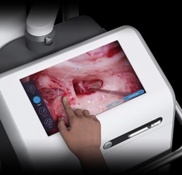
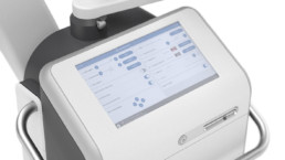
Live support via telestration
Supporting the surgeon with useful information has never been as easy. The touchscreen user interface allows for viewing and telestrating on the surgical field, highlighting e.g. crucial anatomical structures.
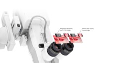
Synchronized surgeon’s view
All information becomes visible right in front of the surgeon’s eyes in the digital binocular. The surgeon remains focused on the procedure without the need to turn away from the binoculars.
Live support via telestration
Supporting the surgeon with useful information has never been as easy. The touchscreen user interface allows for viewing and telestrating on the surgical field, highlighting e.g. crucial anatomical structures.
Synchronized surgeon’s view
All information becomes visible right in front of the surgeon’s eyes in the digital binocular. The surgeon remains focused on the procedure without the need to turn away from the binoculars.
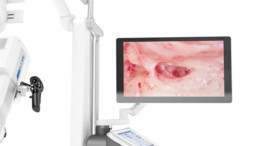
See with the surgeon’s eyes –Share the experience instead of just watching
The ARRISCOPE provides top-of-the-line imaging for a vivid educational experience. The operateur, residents and other observers inside and outside the OR view exactly the same image – the same field of view at the same magnification and focus in an outstanding 3D and 4K image quality.
Further ARRISCOPE 1.1 Highlights
Further ARRISCOPE 1.1 Highlights

- High resolution 3D 4K videos for display of finest details
- The choice between surgery using a high-resolution stereoscopic eyepiece or a 3D 4k monitor
- Videos can be displayed wirelessly on up to four monitors at once
- Video capture can be added with up to 60 seconds of prior imaging, so that significant sequences can be recorded and not lost
- Natural 3D viewing and color reproduction
- 32” integrated 3D 4k monitor for an optimal co-observation experience
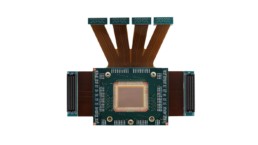
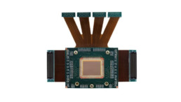
- The core imaging components (camera, illumination and software) developed and produced by ARRI
- Each component’s performance and specification has been fine-tuned towards top-of-the-line image quality
- Oscar® awarded video technology for top-of-the-line surgical imaging
- Efficient cold-light LED illumination prevents tissue from heating while also providing optimal illumination of the surgical field
- The two-way illumination reduces shadows in the image and provides a homogeneously illuminated field of view
- Long lifetime in comparison to conventional xenon illumination

- Intuitive user interface providing access to all system settings
- A dedicated start-up screen guides through the entire setup of the ARRISCOPE before surgery
- The large touchscreen allows for displaying and annotating the live video during the Assist Mode

- The stand allows for effortless one-hand operation and easy positioning of the microscope head over the region of interest
- The ARRISCOPE provides a stable image due to its stand‘s solid mechanical and vibration dampening design
User Testimonials

“When it comes to the requirements for digital ENT microsurgery, ARRI Medical has a focus like almost no other manufacturer. We see this clearly both in routine clinical applications and in research and education. I find ARRI Medical to be a reliable partner for developing new applications in digital ENT microsurgery.”
Prof. Prof. h. c. Dr. med. Thomas Lenarz

“The ARRISCOPE is a huge advantage for our ear surgery courses, because all the students can watch the procedure with previously unheard-of image and detail quality.”
Prof. Dr. J. M. Müller

“Along with routine use, the major advantage of the ARRISCOPE for me is that trainee physicians and clinical staff can share exactly the same view as the surgeon, in 3D and the highest image quality.”
Prof. Dr. Robert Mlynski

“The people at ARRI are specialists. They are highly flexible and react quickly.”
Prof. Dr. med. Dr. h. c. Roland Laszig

“A fantastic tool for every day surgical work.”
Prof. Dr. med. Randolf Riemann
Frequently Asked Questions
The ARRISCOPE is a fully digital surgical microscope which, in contrast to conventional optical systems, displays the situs in a digital binocular with high resolution and in 3D.
The 3D visualization for the surgeon as well as for co-observation and assistance is not an additional purchase option as with conventional systems. Rather, it is immanent in the ARRISCOPE system as digital 3D native imaging is its nature.
The philosophy of the ARRISCOPE is that surgeons remain fully concentrated and do not have to turn away from the situs during the procedure – just like in a surgical cockpit.
Complimentary information can be displayed via PiP Mode (Picture-in-Picture Mode) in outstanding image quality directly in the digital binocular.
The mechanical design allows for almost effortless positioning of the microscope providing a steady image at all times.
The Assist Mode provides live support via telestration. The touchscreen user interface allows for viewing and telestrating on the surgical field displayed, highlighting e.g. crucial anatomical structures. All information becomes visible right in front of the surgeon’s eyes in the digital binocular.
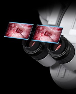
Presentation of supplementary image information as PiP (picture-in-picture) directly in the binocular with maximum image quality. The surgeon can switch between the video sources at any time using the system handles. In principle, signals from a wide variety of external video sources can be displayed. Examples:
Preoperative image data (e.g. CT, MRT)
These can be imported directly from the hospital network (PACS). The surgeon himself can enlarge/reduce the images and scroll through the layers.
Endoscope image
The live video of endoscopes can be displayed directly in the binocular. The PiP display, or in this case even the simultaneous display of the endoscope and microscope image, allows you to “look around the corner” while at the same time seeing the microscope image as a reference for orientation.
The surgeon and co-observer / assistant always see the same 3D image – the same image detail in the same format in comparable quality and always in focus. Co-observers and assistant share exactly the same image and view.
The surgeon can rest assured that the video recordings show exactly what he sees in the eyepiece. There is no difference in terms of field of view, sharpness and focus.
The imaging chain of the ARRISCOPE, consisting of camera (even the chip), LED illumination and corresponding software, is produced by ARRI. All components are coordinated for the best possible image quality. This is in contrast to conventional surgical microscopes whose imaging chain is primarily based on off-the-shelf components.
With the ARRISCOPE cumbersome cabling in the OR becomes obsolete. The ARRISCOPE provides wireless 3D video transmission to up to 4 displays at the same time.
ARRI Medical understands that the OR environment becomes increasingly complex. Therefore, the ARRISCOPE was designed for ease-of-use.
E.g. a dedicated start-up guide leads users through the entire set-up procedure. Important user guidance may be called up at any point of time.
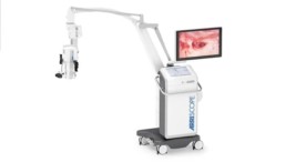
The ARRISCOPE’s small footprint allows easy maneuverability even in small ORs. The long reach of the ARRISCOPE prevents the need for continuous repositioning of the system while still being able to reach any ROI required.
The ARRISCOPE’s LED cold-light source significantly reduces the likeliness of skin burns even at a high light intensity.
Our outstanding service combined with more than 100 years of experience in professional video equipment make ARRI Medical a partner you can always rely on. A proven record of our excellence in video making and imaging is the Oscar we have won for our ALEXA digital camera system.
Technical service:
The ARRISCOPE features remote service for quick system diagnosis and efficient service deployment thus limiting potential down times.
Multiple options allow for tailoring the technical service towards your individual budget and needs – from preventive maintenance to full-coverage services.
Surgical Imaging:
In addition to the surgical microscope ARRISCOPE and all related technical services ARRI Medical allows you:
- Film in the OR: With the more than 100 years of experience of ARRI in the film industry, and its Oscar-winning camera technology, ARRI Medical guarantees professional documentation of your surgery, conference or seminar.
- Post-production: Digital material can be processed according to your wishes. Digital image post-processing, including targeted color adjustment, and sound processing, such as subtitles, audio commentary and dubbing, are possible.
- Conference techology: Professional transmission of live surgeries in large format and 3D at conferences and seminars. Viewers experience an excellent image with the highest quality equipment.
Absolutely! Please contact the ARRI Medical team through the “Contact” button on this website to arrange for a personal visit and demonstration of the ARRISCOPE.
Get in touch
Sign up for information, offers and product demonstrations.
